Analysis and occurrence of fibrous erionite minerals in New Zealand: some technical notes
1. Introduction
Further to the paper presented in NZ Geomechnaics News, Issue 98 in 2019 (Brook et al., 2019), there has been further progress in our research into naturally-occurring zeolitic mineral fibres such as erionite in New Zealand. This article is in response to several requests by geologists and engineers at various consulting companies in New Zealand. Our aims here are two-fold: to provide (1) an update regarding our research, and (2) some guidance about analysis and identification of soils and rock that may contain erionite. From our research findings so far, erionite appears to be rather limited in its distribution and concentration in the Auckland region. Moreover, although our laboratory analysis work in Auckland is still in its infancy after Covid-19 lockdowns, there are few sites that we have found so far where potentially carcinogenic erionite is present. The sites where we have found it, it is in very low concentrations and/or below detection limits in rock. So far, only a few fragmentary erionite fibres have been found transported in the air, and were deposited on vegetation.
To recap, natural zeolites (as opposed to synthetic zeolites) are alteration minerals that form at low temperatures (200°C), from volcanic glass, poorly crystallised clay, and feldspar or silica in wet situations in a variety of sedimentary and volcanic rocks, and in soils (Hay and Sheppard, 2001). More than 60 natural zeolite species are recognised, with most occurring as non-fibrous minerals. However, the zeolites clinoptilolite, dachiardite, edingtonite, erionite, ferrierite, gonnardite, kalborsite, mesolite, mordenite, natrolite, offretite, paranatrolite, scolecite and thomsonite are known to occur as fibrous (“asbestiform”) minerals (Patel et al., 2022, in press). Of these fibrous zeolites, it is only erionite that has been recognized as a Group 1 Carcinogen by the International Agency for Research on Cancer (IARC, 1987). This carcinogenic classification is due to erionite fibres being unequivocally linked to the malignant mesothelioma epidemic in the Cappadocia region of Turkey in the 1970s (Mumpton, 1979). Therefore, identifying where erionite may occur, in what concentrations, and being able to distinguish erionite from other zeolites has important implications for risk assessment of the disturbance of rocks and soils. A further fundamental risk issue is whether any the fibres are actually respirable. For example, inhalable dust is <100 ∝m and this is generally visible and can only affect the upper respiratory tract. However, respirable dust is smaller (<10 ∝m), is invisible under normal conditions, and their extremely small size also means they can be breathed deep into your lungs and lead to lung damage (WorkSafe Queensland, 2020).
In New Zealand, respirable asbestos (probably also appropriate for other mineral fibres such as erionite) is defined by the Health and Safety at Work (Asbestos) Regulations (2016) as an asbestos fibre that:
• is less than 3 µm wide;
• is more than 5 µm long; and
• has a length to width ratio of more than 3:1.
2. Occurrence of Erionite in New Zealand: an update
In the initial 2019 paper that was published, there were significant unknowns then about the occurrence of erionite in New Zealand (i.e., was it “asbestiform”?), and also how erionite may best be analysed and identified. Since then, some progress has been made, and this has been funded by capacity-building grants provided by MBIE and the Royal Society of New Zealand. Within New Zealand, zeolites are found in Northland, Auckland, Coromandel Peninsula, the Taupo Volcanic Zone (TVZ), and Southland (Christie et al., 2002). Erionite has been reported from eighteen locations in New Zealand, in six different regions in the North and South Islands, including multiple locations throughout the Auckland Region.
The main geological occurrences for erionite is volcanic and volcanically-derived rocks that have been subjected to relatively low temperature alteration (Mumpton, 1979). The host rocks or erionite are typically basaltic and rhyolitic tuff. However, erionite has also been identified in sedimentary and metamorphic rocks (Schmieder and Jourdan, 2013; Sheppard, 1996). In sedimentary rock for example, zeolites typically form the cementing matrix, crystallizing within pore spaces, and this can lead to a harder “zeolitized” horizon layer (Figure 1A). This is often more resistant to erosion relative to the surrounding sedimentary layers.

Figure 1: (A) Occurrence of erionite in Waitemata Group Timber Bay Formation, Puketotara peninsula, Kaipara area, with cemented layer at base of ash unit; B) Cemented ash layer that contains disseminated erionite, Timber Bay Formation. Erionite has been detected at 75% in this material. C) Erionite (white) in vesicles within basalt, Waitakere. D) Fibrous zeolites veins (white) in rhyolite, Coromandel Peninsula.
The most commonly reported erionite occurrence is disseminated within ash layers (Figure 1A, 1B), or within vesicles (Figure 1C) and very rarely was erionite reported in a vein (Figure 1D). Within basaltic rocks, erionite tends to form within vesicles (e.g., Wise and Tschernich, 1976), while in felsic rocks, erionite typically occurs disseminated in the matrix of the host rock (Mumpton, 1979), but can also be found to form within vesicles (Barrows, 1980). An example where we have found asbestiform erionite in vesicles is within the Waitakere Group Volcanics at Te Henga Quarry, specifically the Waiatarua Formation (Brook et al. 2022). An example of where we have found erionite as a matrix is in the Waitemata Group Timber Bay Formation at Kaipara (Figure 1A). It must be cautioned though, that we have not found large quantities of erionite at all so far. Rather, erionite seems to be restricted to very restricted geographic locations within rock units. The majority of erionite crystals are very small and are difficult to identify even with a microscope. Erionite fibres can occur individually, or as “bundles”, “radiating clusters”, or a “woolly mass” (Dogan & Dogan, 2008).
3. Technical Analysis of Erionite
Identifying erionite is notoriously difficult for a range of reasons (laboratory equipment, training, lack of accepted standards, disagreements about erionite chemistry etc). Binocular microscopy may be used to determine if fibrous minerals are present within a sample, but it is unreliable when discriminating amongst different mineral fibres. In the USA, the difficulties in identifying erionite were highlighted when unidentified erionite samples were provided for testing transmission electron microscopy (TEM) proficiency for the National Voluntary Laboratory Accreditation Program (NVLAP). Based on this assessment, only 3 out of 76 accredited laboratories correctly identified erionite (Bruce and Strouse, 2009). The morphology of erionite is the primary reason the mineral is toxic (Dogan and Dogan, 2008). Therefore, for the identification of fibrous erionite, using a combination of analytical techniques, including the following as part of a staged approach, is usually recommended: x-ray diffraction (XRD), scanning electron microscope (SEM), with energy dispersive x-ray spectroscopy (EDS) and transmission electron microscopy (TEM) equipped with EDS. These techniques can then analyse the morphology and provide information on the chemical composition. For air samples filters, TEM and SEM-EDS are the most utilised methods, following asbestos reference methods (e.g. NIOSH 7402).
Polarized light microscopy (PLM) has been used occasionally as a screening level analysis, but even in the USA, such analysis of erionite is probably limited to only a few laboratories that have access to high-dispersion refractive index liquids with the appropriate refractive index range (n = 1.460 to 1.480). They would also have needed to have developed a modification to NIOSH Method 9002 to semi-quantitate zeolite fibres (Berry et al. 2019).
Phase contrast microscopy (PCM) has been occasionally used for detecting erionite in soils, an example being those sampled in eastern Montana and western South Dakota (Farcas et al. 2017). For soils, a fluidized bed asbestos segregator (FBAS) can be used to separate the fibres from the soil. This soil preparation method uses air elutriation to separate mineral fibres, such as erionite, from soil particles with higher aerodynamic diameter and deposits those mineral fibres onto filters that can then be analysed using analytical techniques. Previous research on asbestos fibres has demonstrated that low level (0.002% to 0.005% by weight) detection can be readily attained using the FBAS separation method (Farcas et al. 2017). Methods used in the USA to analyse asbestos and other fibres by PCM are covered within NIOSH 7400.
X-Ray Powder Diffraction (XRD) is often used initially in a testing programme to identify the crystallography of the material. This can be used on a single mineral, if enough of the mineral is available, or bulk samples, providing information on the individual minerals that are within a sample. XRD provides spectral data that can be analysed with specialist software. Bulk sample analysis can identify the presence of erionite, identifying other minerals, including zeolites in a given sample. However, erionite and other zeolites can occur together and exhibit very similar XRD patterns, with low concentrations of erionite masked by diffractions from other minerals (Dogan and Dogan, 2008). There are important set-up steps for XRD analysis of putative erionite, including using the side-loading technique, rather than back-loading, so that compaction and bias due to preferential orientation is minimised. Thus, with side-loading, the material falls under gravity into the specimen holder. Second, reducing the scattering of the primary beam is important (can be achieved by narrowing the splits), otherwise “shadowing” effects can occur in the low angle region of the XRD diffractogram, and peaks where erionite would occur are masked. If these steps are not followed carefully, then the XRD approach is pointless in terms of trying to identify the presence of erionite in a sample. Moreover, the minimum amount of mineral needed to be present within the sample is 1–2%, in order to be detected (Meeker, 2008). Therefore, XRD should be used in combination with other analytical techniques.
Scanning Electron Microscopy (SEM) is often used after XRD, to produce images. SEM uses a focused beam of electrons to create a magnified image that can identify erionite mineral fibres as shown in Figure 2A. The images of rock specimens can provide detailed information on the morphology of minerals, particularly useful in the identification of fibrous zeolites such as erionite (Giordani et al., 2017). Sample preparation is straight forward and by scanning over a sample manually, observations should be able to detect erionite-like minerals which can be compared visually with online-images. If the SEM is equipped with EDS (Energy Dispersive Spectroscopy; see below), then chemical composition can also be provided, which assists in mineral identification.
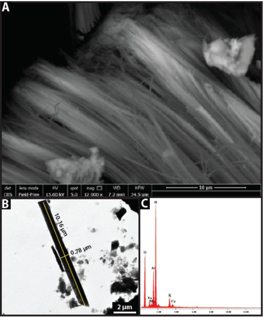
Figure 2: Example data output from Auckland erionite: A) Fibrous erionite from Te Henga Quarry, Auckland; B) TEM image of mineral fibres from Riverhead, Auckland. C). EDS from SEM from a sample from Riverhead, Auckland.
Transmission Electron Microscopy (TEM) is sometimes used as a third stage of analysis. Similar to SEM, it uses an electron beam to produce a magnified image that can identify the internal morphology of erionite mineral fibres, as shown in Figure 2B. In particular, TEM is used to image the morphology of individual mineral fibres of fragile zeolites, such as erionite. This method can determine the length and width of mineral fibres. Many zeolites are sensitive to the electron beam and once the energy of the electron beam collides with the erionite fibres, they deform (Ray, 2020). This degradation caused by the high-energy electron beam (up to 100 kilovolts, kV) can influence the chemistry and crystal structure. Thus, cryogenic holders should be used to protect erionite fibres from the energy of the electron beam (Ray, 2020). Moreover, sample preparation of fibres for analysis can result in over-milling, which destroys the fibre structure (Ray, 2020), biasing any morphological analysis on where the fibres are respirable. Again, if the TEM is equipped with EDS (Energy Dispersive Spectroscopy; see below), then chemical composition can also be provided, which assists in mineral identification. For soils, Berry et al. (2019) used an FBAS apparatus to separate erionite fibres from soils, with nominal concentrations ranging from 0.1% to 0.0001% by weight. The resulting filters were analyzed by TEM, with a method detection limit of 0.003% by weight achieved which is approximately 100 times lower than typical detection limits for soils
by PLM.
Energy Dispersive Spectroscopy (EDS) provides chemical composition of the sample and can be coupled with SEM or TEM imaging, providing spectral data of the elemental compositional as shown in Figure 2C. The EDS is pointed and focused on a selected mineral during SEM or TEM imaging. EDS can be used to help accurately identify between different minerals with similar morphology. The spectrum provides elemental composition of the selected sample, but it should be noted that the electron beam has been found to replace cations in some studies, causing fibres to become unstable, especially if their diameter is <0.5 µm (Ray, 2020).
4. Summary
Erionite can be extremely toxic, and the erionite hazard in New Zealand is still not fully understood, but it appears the risk is mitigated by several factors: (1) erionite appears to be a rare mineral in New Zealand; (2) the temperate maritime climate of NZ contrasts with previous cases of known erionite exposure risks such as central Turkey and North Dakota; moisture possibly mitigates the liberation of erionite fibres from soil and rock into the air; (3) dust management on New Zealand ground engineering sites is already undertaken, and in confined spaces in particular, PPE is already used to mitigate dust inhalation including for silicosis risk. A staged approach (e.g., Figure 3) is probably the most appropriate approach to understand erionite risk, whether or not risk management follows ALARP (as low as reasonably practicable) or SFAIRP (so far as is reasonably practicable; ONRSR, 2021).
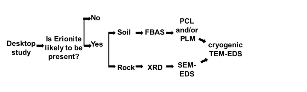
Figure 3: Flow diagram of suggested staged approach to managing erionite hazards.
Further information on the sample type, sample preparation, sample analysis and data output for the analysis of erionite can be found in University of Auckland documents ‘Identification, Sampling and Analysis of Erionite Rock Material: A Technical Note’. The University’s health and safety protocols associated with sampling and analysis of erionite are covered in ‘Erionite Tikanga Akuaku / Health and Safety Protocols’, and either of these documents can be provided by Martin Brook (m.brook@auckland.ac.nz).
References
Berry D, Januch J, Woodbury L, Kent D. (2019). Detection of Erionite and Other Zeolite Fibres in Soil by the Fluidized Bed Preparation Methodology. Microscope 67(4): 147-158.
Barrows KJ. (1980). Zeolitization of Miocene volcaniclastic rocks, southern Desatoya Mountains, Nevada. GSA Bulletin 91, 199–210.
Brook MS, Black PM, Salmond J, Dirks K, Berry T-A, Steinhorn G. (2019). Exposure to erionite: health effects and implications for geotechnical risk management in the New Zealand construction sector. New Zealand Geomechanics 98: 78-81.
Brook MS, Patel J, Di Giuseppe D, Scognamiglio V, Gualtieri A, Kah M, Hamilton A. (2022). Erionite in Auckland, New Zealand: occurrence, character and preliminary assessment. 23rd International Mineralogical Association General Meeting, Lyon, France, 18-22 July.
Bruce S, Strouse GF. (2009). Proficiency testing for achieving accreditation in thermometry. International Journal of Thermophysics 30(1): 351–359.
Christie AB, Brathwaite RL, Thompson BN. (2002). Mineral commodity report 23 – Zeolites. New Zealand Mining 31: 16-24.
Dogan AU, Dogan M. (2008). Re-evaluation and re-classification of erionite series minerals. Environmental Geochemistry and Health 30: 355–366.
Farcas D, Harper M, Januch JW, Jacobs TA, Sarkisian K, Stetler LD, Schwegler-Berry D. (2017). Evaluation of Fluidized Bed Asbestos Segregator to Determine Erionite in Soil. Environmental Earth Sciences 76: 126.
Giordani M, Mattioli M, Dogan M, Dogan AU. (2017). Potential carcinogenic erionite from Lessini Mounts, NE Italy: Morphological, mineralogical and chemical characterization. Journal of Toxicology and Environmental Health, Part A 79: 808–824.
Health and Safety at Work (Asbestos) Regulations, New Zealand Statutes (2016). http://www.legislation.govt.nz/regulation/public/2016/0015/19.0/DLM6729706.html.
IARC (1987). Erionite – IARC Summary & Evaluation, Volume 42.
Hay RL, Sheppard RA. (2001). Occurrence of zeolites in sedimentary rocks: an overview. In: Bish DL, Ming DW (eds), Natural zeolites: occurrence, properties, applications. Reviews in Mineralogy and Geochemistry, volume 45, Mineralogical Society of America, pp. 217-234.
Meeker GP. (2008). Statement of Gregory P. Meeker, Geologist: Asbestos. Retrieved from: https://www.usgs.gov/congressional/statement-gregory-p-meeker-geologist
Mumpton FA. (1979). Mineralogy and Geology of Natural Zeolites. Southern Printing Company.
NIOSH 7400. Asbestos and other fibers by Phase Contrast Microscopy (PCM). Method 7400, Issue 3, June 14, 2019, NIOSH Manual of Analytical Methods.
NIOSH 7402. Asbestos Fibers by Transmission Electron Microscopy (TEM), Method 7402, Issue 2, August 15, 1994, NIOSH Manual of Analytical Methods.
NIOSH 9002. Asbestos (bulk) by Polarized Light Microscopy (PLM). Method 9002, Issue 2, August 15, 1994, NIOSH Manual of Analytical Methods.
ONRSR (2020). ONRSR Guideline – Meaning of duty to ensure safety so far as is reasonably practicable. Office of the National Rail Safety Regulator, Adelaide. 16p.
Ray R. (2020). Discerning Erionite from Other Zeolite Minerals During Analysis. Environmental and Engineering Geoscience 26: 133–139.
Patel J, Brook M, Kah M, Hamilton A. (2022, in press). Global geological occurrence and character of the carcinogenic zeolite mineral, erionite: a review. Frontiers in Chemistry.
Schmieder M, Jourdan F. (2013). The Lappajärvi impact structure (Finland): Age, duration of crater cooling, and implications for early life. Geochimica et Cosmochimica Acta 112: 321–339.
Sheppard RA. (1996). Occurrences of erionite in sedimentary rocks of the Western United States (Open-File Report No. 18), U.S. Geological Survey.
Wise WS, Tschernich RW. (1976). The chemical compositions and origin of the zeolites offretite, erionite, and levyne. American Mineralogist 61: 853–863.
WorkSafe Queensland (2020). Hazardous dusts. Retrieved from: https://www.worksafe.qld.gov.au/safety-and-prevention/hazards/hazardous-exposures/hazardous-dusts#:~:text=Inhalable%20dust%20(smaller%20than%20100,invisible%20under%20normal%20lighting%20conditions.